Anatomy of the retina
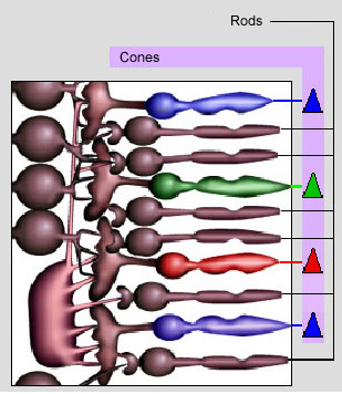
The retina contains light-sensitive cells and other nerve cells, devoted to information processing and transmission of this information up to the brain. The thickness of the retina is about 250µm except around the fovea where it is thinner., Approximately 150 millions of cells are present on the retina, spread over a total area of about 1100 mm2 , divided schematically into three main layers (see figure 3):
1. The layer containing retinal photoreceptors (about 100 million rods and 5 million cones). Rods are highly stretched in one dimension (rod-like shape), and lie perpendicular to the retina surface; they form a dense and regular mosaic.
2. The granular layer containing bipolar cells.
3. The layer containing ganglion cells, which are spiking nerve cells (neurons). They all recombine to form the optic nerve which transmits nerve impulses up to the lateral geniculate nucleus.
![[zoom...]](javascript:window.open(%22../res/Figure3_2.jpg%22,%22_blank%22,%22width=%22+Math.min(800,screen.availWidth)+%22,height=%22+Math.min(600,screen.availWidth)+%22,left=%22+(screen.availWidth-800)/2+%22,top=%22+(screen.availHeight-600)/2+%22,scrollbars=yes,resizable=yes%22)?void(0):void(0))
Photoreceptors: cones and rods
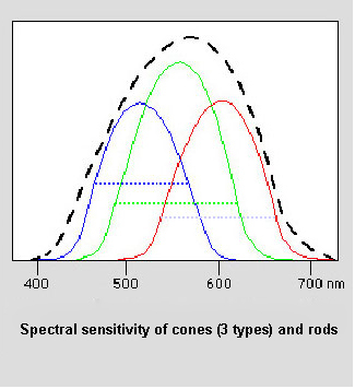
There are 4 different kinds of retinal photoreceptors, three kinds of cones and only one kind of rods. They all have broad sensitivity spectra but with clearly distinct maxima that allows them to be discriminated. These maxima of sensitivity occur at:
-
495 nm for rods
-
420 nm for S cones (Short wavelength or B (blue))
-
530 nm for M cones (Medium wavelength or G (green))
-
560 nm for L cones (Long wavelength or R (red))
The spectral sensitivity curves for cones are given in figure 4. They have similar shapes and overlap significantly.
![[zoom...]](javascript:window.open(%22../res/Figure4_1.jpg%22,%22_blank%22,%22width=%22+Math.min(800,screen.availWidth)+%22,height=%22+Math.min(600,screen.availWidth)+%22,left=%22+(screen.availWidth-800)/2+%22,top=%22+(screen.availHeight-600)/2+%22,scrollbars=yes,resizable=yes%22)?void(0):void(0))
At low light levels (scotopic vision: luminance<103cd/m2), rods work alone. At high levels (photopic vision; luminance>10cd/m2) only cones are active. For intermediate levels (mesopic vision; 10-3 to 10cd/m2) the two types of photoreceptors are involved.
Trichromatic encoding
Since the three types of cones have specific absorption spectra, a given radiation incident on the eye will yield three different and simultaneous physiological responses. This is called trichromatic encoding of color vision: this very important property of human vision is the foundation of the whole theory of colorimetry. The trichromatic theory was first proposed by Young in 1801, further explored by Helmholtz in 1852, and was later confirmed by micro-spectrophotometric experiments [] as well as by electrophysiological measurements [].
However, the existence of three different types of cones does not allow explaining all the features of color vision. There are many physio-psychological aspects that play a role in the whole process of color vision, which are still subject of intensive research. Colorimetry deals only with the interaction of light with the photosensitive cells of the retina.